Visual health

Cornea & lens

The cornea is an important component of the eye's optical system, which is why it has become the preferred site of action for various refractive surgery techniques that aim to modify its optical power by reshaping its anterior surface.

The cornea’s defining feature is its transparency: it is avascular, has no pigment, its nerves lack myelin, and its stroma has a regularly arranged collagen fiber structure.

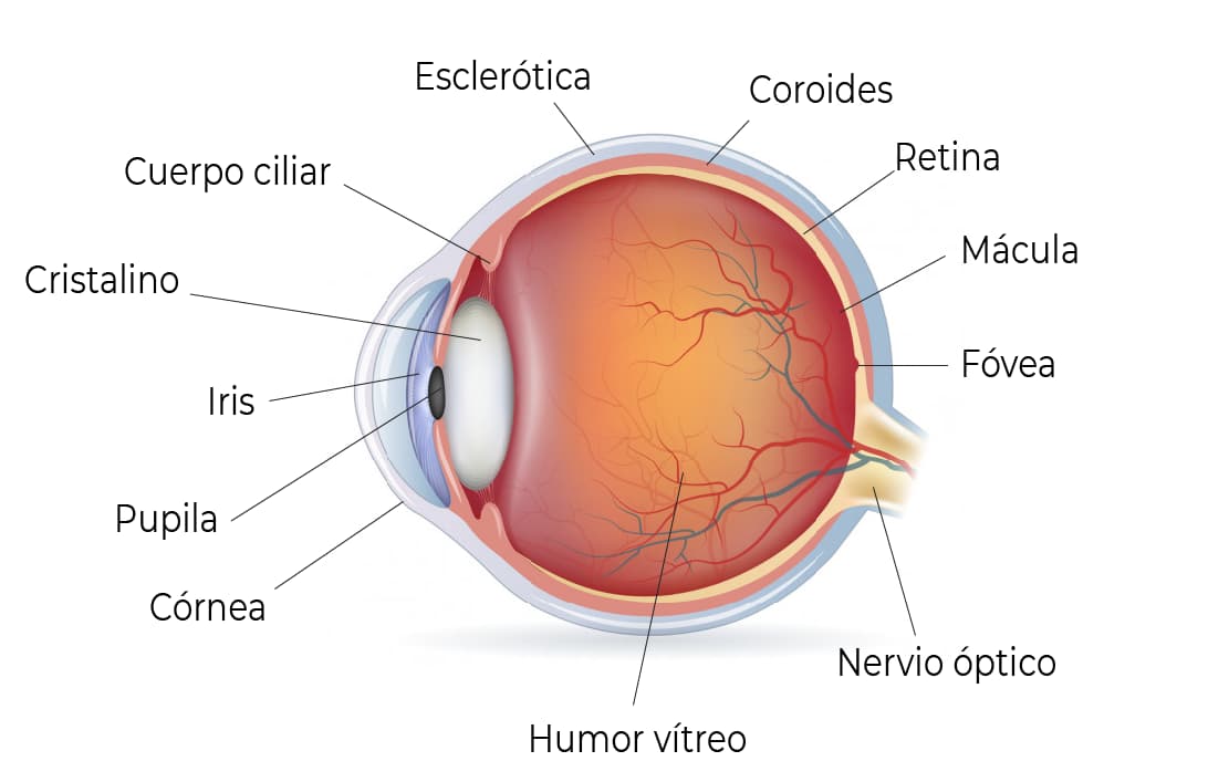
Anatomy of the eye and its parts

Corneal disorders

Keratitis

Inflammatory and infectious processes of the cornea are grouped under the term keratitis; when the conjunctiva is affected as well, it is called keratoconjunctivitis. These are generally focal defects of the corneal epithelium, and are sometimes asymptomatic.

Corneal ulcers

The corneal stroma is generally affected, but this is preceded by an epithelial alteration.

All ulcers present with acute pain, ciliary hyperemia, decreased visual acuity, tearing and photophobia.

Keratoconus

Keratoconus literally means a cone-shaped cornea. It is a progressive, uncommon disease in which the cornea becomes abnormally thin and bulges forward.

Blurred vision and image distortion are the first symptoms, usually appearing in the late teens to early twenties. In the earliest stages, vision may be only slightly affected, causing glare, light sensitivity and irritation.

Dystrophies and degenerations

These are a group of primary corneal disorders, generally hereditary and bilateral, that show no signs of inflammation or neovascularization and, in some cases, are associated with systemic diseases. The treatment for the most severe cases is corneal transplant.

Lens

The lens sits behind the iris, encapsulated and held to the ciliary body by the zonular fibers. It is a biconvex lens whose main function is to focus images at different distances — a process also called accommodation.

Like the cornea, this structure is characterized by its transparency: both are avascular, have no pigment, their nerves lack myelin, and their stroma has a regularly arranged collagen fiber structure.

Anatomy of the eye and its parts

Cataracts

This is the loss of transparency and mobility of the lens. Cataracts can appear at any age, but tend to be more common in people over 60 due to the passage of time. Congenital cataracts occur in children born with them due to hereditary factors or illnesses during pregnancy; others develop cataracts early due to metabolic diseases. They can also be associated with severe myopia and eye trauma.

Over the years, or due to some illness, the lens loses its natural transparency and elasticity and becomes an opaque, rigid lens, losing its function in the process. A cataract can be mild or advanced depending on how much transparency the lens has lost. Visual quality is affected in proportion to how advanced the cataract is.

Treatment

Treatment is surgical. The most modern technique is called phacoemulsification, and consists of removing the lens using ultrasound; it is then replaced with an intraocular lens placed in the same location, allowing the person, in most cases, to recover their lost vision.

It is an outpatient surgery, performed under local (eye-drop) anesthesia, with few complications. Patients are generally able to return to their activities within a short period of time. There are currently several models of intraocular lenses or artificial implants available.

Want to get in touch with us?

You can call or message us on WhatsApp at +549 11-5823-7463 (WhatsApp and calls), or call +549 11-5823-2375 (calls only).